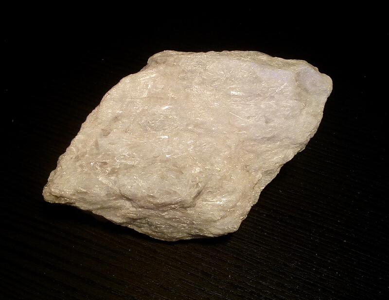
SC301 - Anthophyllite, Clino-suenoite, Tremolite
Locality: International Talc Company Mine (Reynolds Mine), Talcville, St. Lawrence Co., New York, USA (MINDAT)
Size: 10 x 6 x 2 cm - Cabinet (6-10 cm)
Weight: 145 g
minID: 7H1-1UY
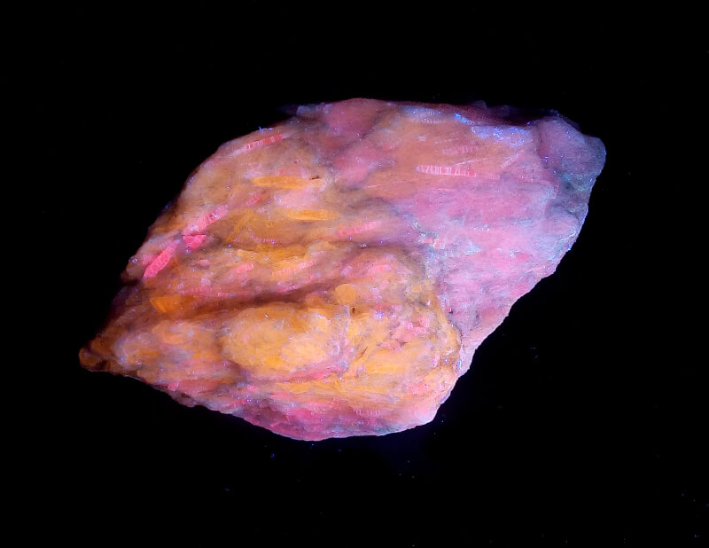
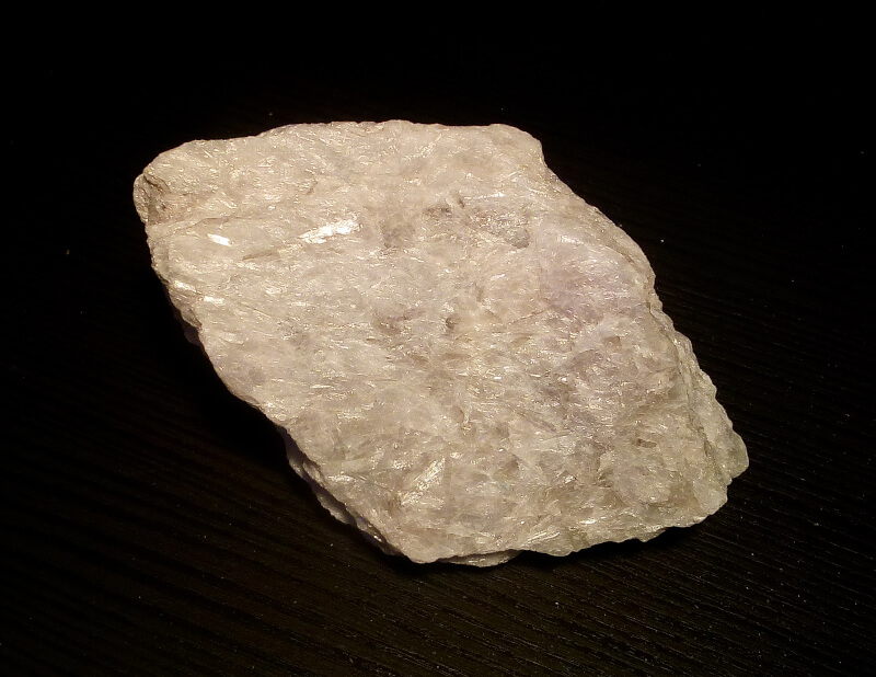
Tremolite mixed with “tirodite”, i.e. anthopyllite and/or clino-suenoite. Under long wave UV light the tremolite fluoresces yellow/orange while the antophyllite/clino-suenoite fluoresces red/violet. Under shortwave UV light or 405nm laser the red/violet essentially disappears and the whole specimen fluoresces yellow/orange. As far as I know it is not possible to distinguish the anthopyllite from the clino-suenoite without elemental analysis.
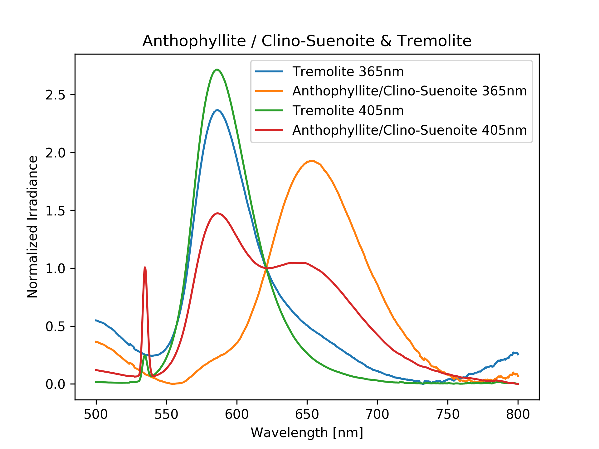
I acquired four fluorescence spectra: two for the tremolite and two for the “tirodite”, under 365nm LED and 405nm blue laser. Both tremolite spectra have the same peak at about about 580nm. This should be due to Mn2+ substituting Ca2+. The spectrum of the tirodite under 365nm led shows a peak at about 650nm (Mn2+ substituting Mg2+ ?), with a shoulder at about 580nm. With the 405nm blue laser the spectrum of the tirodite shows two clear peaks: one at 580nm and one at 650nm. My guess is that the red amphibole spectrum is a mix of the emission of the two minerals: tremolite emitting at 580nm and the tirodite at 650nm. The tirodite fluoresces weaker under shortwave UV light or 405nm blue laser, leaving only the tremolite visible. The intensities in the spectra are normalized and cannot be compared. The sharp peak 535nm is an artifact from the blue laser.